A good friend of mine, Nicole, has asked a great question about the post Sticks and Stones: Basics of Skeletal Trauma.
She says: “Love this! Also if this is the superior view would you say that the trauma occurred along any suture lines? Also out of curiosity, which bone of the skull is pictured?”
Thanks for the question, Nicole! This gives me a great opportunity to talk a little bit about the anatomy of the skull.
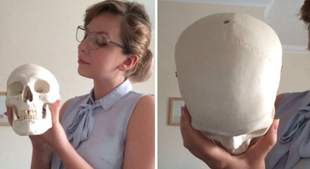
As the original picture only shows a bit of the skull, I’ll bring in my lovely assistant!


You can see that this image of Stan’s head corresponds to the left side of the image above Nicole’s question. I’ll just take the top of Stan’s head off to make it easier to handle.


Nicole wants to know if the trauma on the first skull involved these sutures, and which bones of the skull are pictured.
The bones of the superior aspect of the skull are as follows:


The angle of the trauma to the first skull looks approximately this. The frontal bone is now facing upwards.

As you can see, there are two sutures involved. The vertical suture, called the saggital suture, is cut transversely. The horizontal suture, called the coronal suture, is impacted on the right.
But why are the sutures visible on Stan and not the first skull? Well there are two options.
As we get older the sutures fuse, but it doesn’t just end there! Often the sutures will begin to fade and become obliterated. Sometimes anthropologists will use the level of fading to establish age at death. Buuuuut they shouldn’t. Sometimes the sutures don’t fade at all, and they certainly never fade on a predictable timeline.
The second option might be that the surface of the bone is poorly preserved.
As I don’t currently have access to the bone in question, I can’t confirm, but looking at the picture I suspect it’s a mix of both.
Thanks Nicole!

Leave a comment